COVID-19 – “The Sonographers Dilemma”


COVID-19 – “The Sonographers Dilemma”

This is a personal account of my feelings, thoughts and learnings as a Sonographer/Tutor Sonographer in a Private Practice setting in Australia!

Are your anxiety levels running high?  Mine are!

I’m worried about my elderly Grandparents (90 and 92), my parents, and all of those friends and family in the community who have weaker immunity.

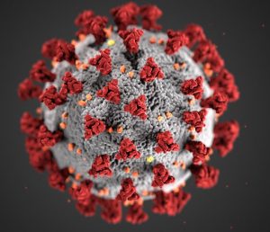
How is this Corona Virus Pandemic going to play out in your region?

How do we as Sonographers help contain the spread, and keep ourselves safe when we are UNABLE to  maintain “Social distancing”.  As Sonographers our patients are close, and we are often in the room with them for 20-30 minutes at a time.

Currently in Australia where I am based, confirmed numbers are relatively low.  But anxiety levels are excessive.  My question especially as a Sonographer in a Private Practice setting, is how do we keep ourselves safe/limit our exposure, to the active virus?  How do we ensure that we don’t contribute to the spread? To our patients and our loved ones?

Currently we are following the Australian Government Department of Health Guidelines (https://www.health.gov.au/)

  • People who have been overseas in the past 14 days are required to self-isolate and will not be examined (can postpone appointment)
  • People who have come into contact with a confirmed case of COVID-19 are also required to self-isolate for 14 days and will not be examined (can postpone appointment)
  • People who have a fever are also asked to re-schedule their appointment.

But what about those people who may have contracted the virus via community transmission (unknowingly) and are asymptomatic.  These are the people we sonographers my encounter.

I watched the ISUOG webinar on the 17/03/2020  and want to share “ my personal key learnings” from the speakers.  The entire webinar can be found at:

Prof Francesco Castelli (Italy), accentuated how quickly the virus spreads, and that it is highly infectious amongst health workers.   He also said that often first testing of the virus can provide a False negative reading…. So be cautious.  If clinical symptoms fit, then treat as COVID-19 as precaution.  He also said that once testing positive, it may take up to 3 weeks for a patient to be non-infectious (not be contagious).

Dr Jill Lee Cheng Sim (Singapore), discuss how the Singaporean health care system had learnt from the SARS outbreak in 2009, where 25% of health care workers were infected.  To date on 1% of health care workers have been infected with the COVID-19 virus.

This is attributed to:

  • Protective equipment and good infection control training.
  • General preventative measures and PPE use.

Also remember to support colleagues, be aware of Mental and social health.

Prof Liona Poon (HK) stated that they wear a mask at all times whilst at work and that even at lunch personal hygiene etiquette (Social Distancing) should be maintained.  This is how health care workers in Wuhan enabled the disease unknowingly.  The appropriate masks have been stated as N2/N95.  That staff in Hong Kong also change their clothes and shower prior to coming home.  Preventing any spread via clothing.

A guide to the correct use of the N2/N95 mask can be found at this site:

https://www.health.qld.gov.au/clinical-practice/guidelines-procedures/diseases-infection/infection-prevention/transmission-precautions/p2n95-mask

I have started implementing my learnings from Tuesdays nights Webinar immediately.  Here is a photo of me with my new fashion accessory  “N2 mask” (Getting use to the feeling on my face)….. trying not to hyperventilate😊

I am continuing with all my usual behaviours also.  Gloving for all patients, cleaning the room and probes thoroughly between patients.

Changed my clothes prior to leaving work and showered as soon as I got home!

We all need to stick together at these tough times.  Stay safe.

Please feel free to touch base with questions/discussion😊

Gail

Director/Tutor Sonographer/Sonographer (Integrated Ultrasound Education)

Email: gail@iuc.consulting.com.au


Aortic Dissection – Not something you see every day!


Aortic Dissection – Not something you see every day!

When you are asked to perform an Abdominal Ultrasound examination, with vague clinical history = “Ongoing back/loin pain for 10 years”.

My first thought is pathology such as: ? Kidney pathology ? Genuine Back pain.  NOT Aortic dissection.


What causes an Aortic Dissection?

The Aorta is one of the major blood vessels that transports blood around the body.  It’s wall is made up of three layers:

  • Tunica intima, tunica media and tunic adventitia.

A dissection occurs with a tear in the intima.  Blood enters this space and tears the intima layer away from the media layer, in effect generating a false channel/lumen.

In some life-threatening cases, it can cause a rupture in all three layers.  Alternatively, if it only affects one or two layers, resulting in generalised back/flank pain can be felt and is treatable if detected early.

True cause is unknown, however Aortic dissections generally occur in:

  • Males aged 50-70.
  • > high blood pressure
  • Conditions such as Marfans Syndrome, Ehlers-Danlos Syndrome, Turners Syndrome.
  • Rare complication of Third Trimester pregnancy and post-partum.
  • Blunt trauma.
  • Complication following surgery i.e. Mitral valve replacement.
  • Use of cocaine.

Following the Stanford classification there is two types of Dissections:

  • Type A which involves the ascending aorta and arch – Surgical Management required.
  • Type B involves the descending aorta – Medical management and blood pressure monitoring.
  • Patients can also have a combination of both – This was the case with my particular patient.

Symptoms:

Generally, symptoms can be similar to that of a heart attack, however specifically the following are indicators:

  • Chest/back pain (Sever)
  • Sudden onset of abdominal pain (Sever)
  • Abnormal blood pressures/change in pulses between arms
  • Loss of consciousness.
  • Leg pain/difficulty walking
  • Shortness/loss of breath

Ultrasound appearances on Ultrasound:

  • Intimal flap on Ultrasound, has both a high sensitivity and specificity for a dissection.

Ultrasound being portable, rapid and easily accessible makes initially diagnosis ideal.

Optimal Imaging for correct diagnosis:

  • Plain Chest Xray (CXR) – Aortic Dissection can be overlooked on a CXR in 12-18% of cases.
  • Arterial enhanced contract CT (CTA). Gold Standard.
  • Multi-planar transesophageal echocardiography.
  • Rapid non contrast MRI (Good for review, but maybe utilised more in acute evaluation).
  • Conventional digital subtraction Angiography (DSA) (However generally replaced by CTA).

Treatment:

  • Surgery – Removal of the dissected aorta or blocking of the channel where blood is entering into the aortic wall is performed. Reconstruction is made with synthetic grafts/stents/tubing, and mesh.
  • Medication – Medication is used to lower blood pressure and prevent the dissection from getting worse.
  • Follow up treatment – Generally lifelong blood pressure medication and ongoing CTA/MRI for review.

References:

‘The Pancreas”


“The Pancreas”…..

Did you know November is Pancreatic Cancer awareness month?

The Cancer Council of Australia states that Pancreatic Cancer is the 5th most common cancer, with an incidence of 1 in 54 for men and 1 in 70 for women.

Unfortunately, the 5 year survival rate is on 8.7%.

(https://www.cancer.org.au/about-cancer/types-of-cancer/pancreatic-cancer.html)

Currently there is no specific screening test for Pancreatic Cancer, which results in late detection of the cancer and thus a poor outcome.

There are commonly 2 types of pancreatic carcinoma’s:

  • Pancreatic Exocrine Carcinoma’s (93% of Pancreatic Carcinomas).
    • Adenocarcinoma being the most common (It develops in the cells lining the pancreatic duct), and most commonly identified in the head of the pancreas.
  • Pancreatic Neuroendocrine Carcinoma’s (7% of Pancreatic Carcinomas).  These develop from abnormal growth of islet cells within the pancreas.                                            (https://www.pancan.org/facing-pancreatic-cancer/about-pancreatic-cancer/types-of-pancreatic-cancer/)

Symptoms that can be associated with Pancreatic Cancer include:

  • Upper abdominal pain
  • Loss of appetite/weight loss
  • Altered bowel habits
  • Jaundice
  • Diabetes

Ultrasound is a fantastic tool for imaging the Pancreas, in a patient that has been correctly prepped.

  • Fasted for 6-8 hours (No eating, chewing or smoking).
  • Utilise a curved ultrasound probe ideally 2-10 Mhz.


Techniques to optimise visualisation include:

  • Deep inspiration/expiration.
  • Distention of the abdomen (Valsalva).
  • Give the patient 2 glasses of water (The water acts as a window).
  • Examine the pancreas in the erect position.


Written by Gail Crawford (Tutor Sonographer/Director IUE).