Clean, Clean, Clean – Minimise Risk, Minimise Exposure!
Clean, Clean, Clean – Minimise Risk, Minimise Exposure, “Social Distance”…. this is all I have heard for the past two weeks. But still as Sonographers we can’t “Socially Distance”.
Everyone is implementing strict guidelines in the Health Profession as to who can be seen/assessed and who should be rebooked at a later date following a quarantine period. Private practices that I am involved with now only allow the patient in the room, unless a carer or guardian is required (limiting any unnecessary exposure from a third person). This includes obstetric patients, so sadly no partners are allowed. Facetime is being implemented at some locations for partner interaction😊
Sonographers in the hospital settings are being regularly updated with strict protocols around dealing with COVID-19 positive or suspected positive patients. Appropriate PPE is being provided, along with detailed cleaning instructions, scan times etc.
But what I wanted to focus on was looking after ourselves “The Sonographer” and those other health practitioners that are in close contact with a patient for extended periods of time, in the LOW risk setting (i.e. no current indication of COVID-19 exposure).
Patients will continue needing our essential ultrasound services. Pregnant women will need their routine screening, we will be needed to exclude ectopic pregnancies, DVTs and appendicitis. Acute Cholecystitis cases will still need our attention. We might see a drop in Musculoskeletal as sporting activities are limited, but may in time to come, need to perform chest Ultrasounds for suspected pneumonia.
How can we minimise our risk and exposure? This is a fast-moving entity. Some key learnings that have been consolidated are:
|
COVID 19 (Officially – SARS-CoV-2) · Can be airborne for up to 3 hours. · Can remain active on hard/shiny surfaces for up to 72 hours · Can remain traceable on porous surfaces(Cardboard/Paper/Fabrics) for up to 24 hours |
Work Attire:
- If you don’t have scrubs, or a uniform. Consider some easy-care cotton clothing (used solely for work).
- Ensure sleeves are short (to ensure optimal hand washing techniques).
- Remove uniform at work, bag and bring home (could bag in pillowcase). Everything into the wash immediately. Hot wash with detergent is recommended.
- Shoes (closed in specific for work). Leave at the clinic/or at the front door before entering the home, reducing the risk of the virus entering the home.
Hand Cleanliness:
- We are all aware that hand cleanliness is essential; it goes without saying…. Gloves at all times. Ensure hand washing techniques with soap and water are up to date/hand sanitiser is utilised appropriately.
Ultrasound room:
- Remove all unnecessary items from the room.
- No bed linen to be used, with beds to be cleaned after every patient.
- Ultrasound Machine and probes to be cleaned after every patient.
- Clean the door handles and desk/ keyboard regularly through the day.
Please review the cleaning products you are using:
- ‘Detergents
- Acidic, neutral or alkaline. For most general cleaning tasks, a neutral detergent with pH between 6 and 8 should be used.
- Disinfectants
- Alcohol wipes with 70-90% alcohol (ethyl alcohol or isopropyl alcohol)
- Chlorine and chlorine compounds- i.e. sodium hypochlorite (household bleach), sodium dichloroisocyanurate (NaDCC) and calcium hypochlorite (bleaching powder)
- Hydrogen peroxide
- Quaternary ammonium compounds (alkyl dimethyl benzyl ammonium chlorides)
- Phenolic disinfectants’ (https://ww2.health.wa.gov.au/~/media/Files/Corporate/general%20documents/Infectious%20diseases/PDF/Coronavirus/COVID19-Environmental-Cleaning-for-workplaces.pdf)
Word of Warning………
- Ensure cleaning products kill viruses not just “Bacteria”.
- Refer to your specific Ultrasound vendor in relation to probe cleaning (As probes can be sensitive to some cleaning products). All vendors have released appropriate cleaning products to combat COVID-19.
|
US Environmental Protection agent list of disinfectants for use against SARS-CoV-2, can be found at his website: https://www.epa.gov/pesticide-registration/list-n-disinfectants-use-against-sars-cov-2 |
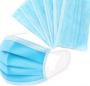
Face Masks (Appropriate PPE):
As sonographers we can’t socially distance. I know a lot of sonographers in LOW risk clinics are concerned about PPE, especially face masks.
We are all aware that correct specification face masks are in short supply – N2/N95 masks (95% effective). – PLEASE ensure that if using these, please use correctly and maintain a full seal.
Dr Jill Lee Cheng Sim (Singapore), also suggested as a barrier “3 ply masks”, for low risk personal (these don’t have a complete seal and thus tiny particles can get through). But it is a good baseline.
Alternatively, due to short supply I have been researching “cloth masks”. My mother has kindly made a prototype and currently making more for my Sonographer friends and colleagues. There is very limited research on the topic and with such a fast paced virus pandemic, not a great deal of time to trial. From a research paper in 2013 Davies et al conclusion was “Our findings suggest that a homemade mask should only be considered as a last resort to prevent droplet transmission from infected individuals, but it would be better than no protection”. https://www.ncbi.nlm.nih.gov/pubmed/24229526.
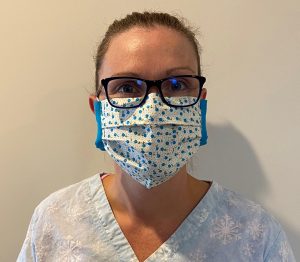
“In a low risk setting I think this is definitely feasible”
Lunch/Tea Room:
- Avoid sharing cups/ mugs in the tearoom. Take your own keep cup if you use the staff room and avoid the communal tea towels etc. Always maintain personal hygiene etiquette (Social Distancing). As this is how health care workers in Wuhan enabled the disease unknowingly.
- Also pay attention to frequently used areas such as toilets/bathrooms, light switches/door handles. Use appropriate hand washing/hand sanitising techniques.
Minimise examination time:
- Scans should be performed efficiently to reduce patient contact time (but by no means does this mean reducing the quality of the examination). This means if a bladder is 3/4 full for a renal, scan them as they present. If the scan requires further prep to be diagnostic, rebook them. Do not leave patients in the room or department to fill. For obstetrics scans, send the patient offsite for a walk and then rescan.
Mental Health:
- While the entire world is grappling to contain the coronavirus, there is a huge emotional and physiological side to this virus. Please everyone be mindful of your mental health. Continue talking to your peers about your concerns. Let’s work at a team to get through this together. Keep up your physical health (exercise is a fantastic stress release), take up a hobby, stay connected with friends and family and remember this is only temporary. We CAN and WILL get through this.
We all need to stick together at these tough times. Stay safe.
Please feel free to touch base with questions/discussion or if you would like a homemade mask (Just as a low risk temporary measure)😊
Gail
Director/Tutor Sonographer/Sonographer
Integrated Ultrasound Education
gail@iuc.consulting.com.au
![]()
Other articles which may be of interest:
- “ISUOG Safety Committee Position Statement: safe performance of obstetric and gynaecological scans and equipment cleaning in the context of COVID-19”. https://www.isuog.org/uploads/assets/d03798de-11ff-4037-beecc9c1495d9572/e6f65fb1-f6af-4d94-beb02bb4ea78c0cc/ISUOG- Safety-Committee-statement-COVID19.pdf
- “Ultrasound Transducer Decontamination – Best Practice Summary”. https://www.sor.org/sites/default/files/images/best_practice_summary_-_probe_decontamination_-_final_13_02_2020.pdf
References:
- Davies A, Thompson KA, Giri K, Kafatos G, Walker J, BennettA. Testing the efficacy of homemade masks: would they protect in an influenza pandemic? Disaster Med Public Health Prep.2013 Aug;7(4):413-8. doi: 10.1017/dmp.2013.43.
- Neeltje van Doremalen, Trenton Bushmaker, Dylan H. Morris, Myndi G. Holbrook, Amandine Gamble, Brandi N. Williamson, Azaibi Tamin, Jennifer L. Harcourt, Natalie J. Thornburg, Susan I. Gerber, James O. Lloyd-Smith, Emmie de Wit, Vincent J. Munster. Aerosol and Surface Stability of SARS-CoV -2 as Comparted with SARS-CoV-1. New England Journal of Medicine, 2020; DOI: 1056/NEJMc2004973
- National Institute of Allergy and Infectious Diseases’ Laboratory of Virology in the Division of Intramural Research in Hamilton, New England Journal of Medicine, 2020
- https://www.huffingtonpost.com.au/entry/how-long-coronavirus-live-clothing-washing_l_5e724927c5b6eab779409e74
- https://ww2.health.wa.gov.au/~/media/Files/Corporate/general%20documents/Infectious%20diseases/PDF/Coronavirus/COVID19-Environmental-Cleaning-for-workplaces.pdf
- https://www.epa.gov/pesticide-registration/list-n-disinfectants-use-against-sars-cov-2
- https://www.health.qld.gov.au/clinical-practice/guidelines-procedures/diseases-infection/infection-prevention/transmission-precautions/p2n95-mask